Wei-Chen CHU (朱韋臣), Ph.D.
- R&D Scientist
ICOB Imaging Core, Academia Sinica, Taiwan - E-mail: weichen01@as.edu.tw
- LinkedIn: https://www.linkedin.com/in/weichen-chu/
- Bluesky: @weichen01.bsky.social
- X (Twitter): @WeiChenCHU1
Profile
- I'm an Imaging Core Facility Manager and Bioimage Analyst passionate about empowering researchers to generate high-quality imaging data and develop reproducible analysis workflows. I teach confocal microscopy and bioimage analysis with FIJI and AI tools, equipping trainees with the skills to independently plan, acquire, and troubleshoot their experiments. As part of my commitment to open science, I develop and share open-access training materials. Beyond training, I design and implement custom bioimage analysis workflows and provide expert consultation for advanced imaging projects.
- If you’re interested in collaboration, or consulting on bioimaging analysis projects, I’d love to connect.🙂
Professional Experience
-
Imaging Core Facility, Institute of Cellular and Organismic Biology (ICOB), Academia Sinica, Taiwan
- R&D Scientist (Jan 2024 - Present)
- Project Scientist (Oct 2021 - Dec 2023)
- Research Technical Employee (Sep 2021)
- Postdocroal Researcher (May 2021- Aug 2021)
-
RIKEN Center for Biosystems Dynamics Research (BDR), Japan
- Research Scientist (Sep 2017 - Mar 2021)
- Visiting Scientist (Oct 2016 - Sep 2017)
Supported by Postdoctoral Research Abroad Program, Ministry of Science and Technology, Taiwan - Research Scientist (Jul 2015 - Sep 2016)
Education
- Ph.D., Life Science
Graduate Institute of Life Science, National Defense Medical Center, Taiwan - Master's, Life Science
Department of Life Science, National Chung Hsing University, Taiwan - Bachelor's, Life Science
Department of Life Science, National Chung Hsing University, Taiwan
Core Skills
- Confocal Microscopy/ Fluorescence microscopy
- Immunofluorescence, fluorescence protein tagging
- Bio-Image processing and analysis using FIJI/ ImageJ, Napari and Imaris
- GPU-accelerated image processing libraries (CLIJ series in FIJI, clEsperanto in Python)
- Developing custom image processing and analysis workflows using ImageJ Macro or Python
Honors & Awards
- Outstanding Employee 2024, Academia Sinica, Taiwan
(113年中央研究院工作績優人員) - Drosophila Image Award 2022: Honorable Mention
- RIKEN Incentive Project FY2018
- Postdoctoral Research Abroad Program 2016, Ministry of Science and Technology, Taiwan
Membership Affiliations
- EABIAS: East Asia Bioimage Analysts’ Society
- Founding member
- Website maintainer
- GloBIAS: Global BioImage Analysts' Society
- Ordinary member
- QUAREP-LiMi: Quality Assessment and Reproducibility for Instruments & Images in Light Microscopy
- Member
Organizing Bioimage Analysis Traing Course
-
ICOB imaging Core summer imaging workshops (2021-present)
-
EABIAS Training event:
Attended Bioimaing and Bioimage Analysis Training Course
-
In-person attendance:
-
Global BioImaging Train-the-Trainer 2025 Course
- Supported by Global Bioimaging Travel Grant
-
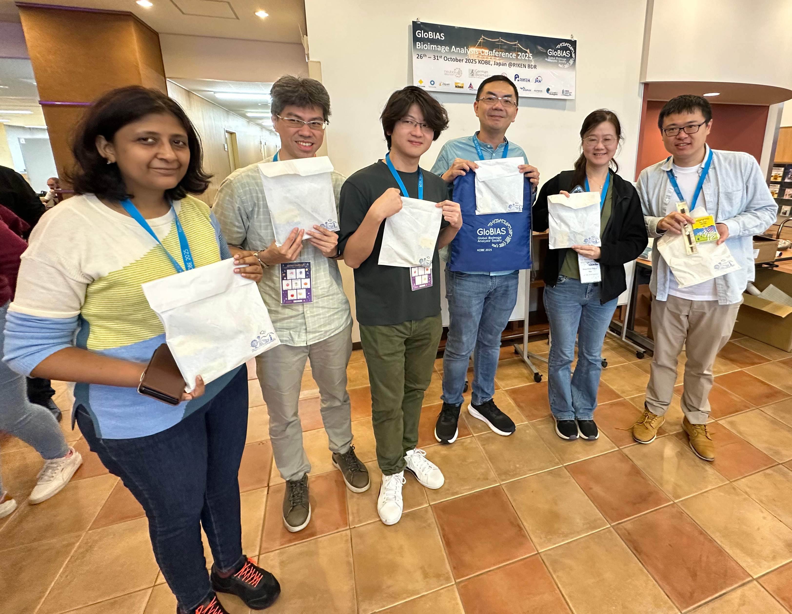
GloBIAS Bioimage Analysis Training School 2025
- Selected trainee, travel supported by ICOB, Academia Sinica, Taiwan
- Award for the projection presentation (Group work with Shao-Chun Peggy HSU, Ji-Ying HUANG, Jen-Chien CHANG, Yung-Li CHEN, and Saumya AGRAWAL)
-
PoL Bio-Image Analysis Training School 2023 (Early Career Track)
- Selected trainee, travel supported by ICOB, Academia Sinica, Taiwan
- NEUBIAS-Style Training School, TU Dresden, Germany
- Bioimage Analysis Workshop 2023 @ICOB
- Python-based bioimage analysis workshop
- Trainee, Assistant and Event Organizer (with Dr. Jung-Kun WEN)
-
-
Online attendance:
-
Introduction to napari workshop (2026)
Pilot virtual introductory workshop on napari, organized by the napari core team. -
YMIA Python-Based Event Series 2024
(Young Microscopists and Image Analysts, German Bioimaging) -
VMCF Bioimage Analysis and Data Processing Workshop 2024-2025
(Viničná Microscopy Core Facility, Charles University, Czechia) -
BOMP Fundamentals of Fluorescence Microscopy 2024-2026
(Biological Optical Microscopy Platform, University of Melbourne)
-
Open-Access Training Materials
-
Practical AI Tools for Bioimage Segmentation: ilastik, Cellpose, and μSAM
- Slide in English (mostly): https://zenodo.org/records/19911180
- YouTube Recording (Mandarin): https://youtu.be/oUlHOoAfHrI
-
Bioimage Analysis with FIJI and AI tools (@ICOB Summer Bioimaging Workshop 2025)
- Slide in English: https://doi.org/10.5281/zenodo.15910878
- YouTube Recording (Mandarin): https://youtu.be/qE78Yqv3UxI
-
Bioimage Analysis with FIJI and AI tools (@TIGP‐INS Neuro‐imaging workshop 2025)
- Slide in English: https://doi.org/10.5281/zenodo.15588682
- YouTube Recording (English): https://youtu.be/nVYhvsN7Jyg
-
Interactive Bioimage Analysis Workflow with CLIJ (@EABIAS 2025 training event)
- Slide in English https://doi.org/10.5281/zenodo.15070246
- YouTube Recording (Mandarin): https://youtu.be/uheSMSENnzE
-
Introducing to Licensing for Documents and Code (@EABIAS 2025 training event)
- Slide in English: GitHub links
- YouTube Recording (Mandarin): https://youtu.be/iGVps1Qazmo?list=PL_9oCBBWdG8mLVflK-MJ3YkUPmhwpKO1s&t=4945
-
Open source AI Tools for bioimage analysis workshop (@ICOB Summer Bioimaging Workshop 2024)
- Slide in English: https://doi.org/10.5281/zenodo.13284351
- YouTube Recording (Mandarin): https://youtu.be/KqwssouW0G0
-
Bioimage Analysis with FIJI /ImageJ & Friends workshop (@ICOB Summer Bioimaging Workshop 2024)
- Slide in English: https://doi.org/10.5281/zenodo.12803966
- YouTube Recording (Mandarin): https://youtu.be/rMRV2N81fkM
-
Bioimage Analysis with FIJI /ImageJ Workshop (@ICOB Summer Bioimaging Workshop 2023)
- Slide in English: https://doi.org/10.5281/zenodo.12736727
- YouTube Recording (Mandarin): https://youtu.be/cnfW9vPQ_XI
Open-Source Bioimaging Analysis Workflows / Tools
-
LLSM-Batch-Preprocessing
This repository contains customized ImageJ/Fiji macros designed for the preprocessing of large-volume Light-Sheet Microscopy raw image data. These scripts utilize GPU-accelerated processing to suppress background noise, enhance the cellular nuclei signal-to-noise ratio (SNR) for spot detection, and smooth structural boundaries for anatomical segmentation, preparing the data for downstream quantification in Imaris. -
yeast-nuclear-puncta-cell-counter
A image analysis pipeline for automated detection and quantification of nuclear puncta in yeast fluorescence microscopy images. -
TF-Nuclear-Translocation-Analysis
This repository provides Python scripts for refining single-cell trajectories and quantitatively measuring stress-induced nuclear translocation of the Dot6–GFP transcription factor from time-lapse microscopy data. The scripts operate after image acquisition, segmentation, and tracking, which are assumed to be performed in Fiji/ImageJ with Trackmate-Cellpose. -
TIRF_vesicle_colocalize_analysis
This repository contains Python code for analyzing time series TIRF (Total Internal Reflection Fluorescence) microscopy images to identify and track colocalized Glut10 and Rab5 vesicles. -
Cell_Dist_Mesh_Generator
This repository hosts a FIJI/ImageJ macro tailored for the automated generation of distance meshes between cells, aiding in quantitative visualization. -
central-nuclei-muscle-analyzer
A comprehensive ImageJ macro for automated analysis of nuclear localization in muscle cross-sectional areas, specifically designed to identify and quantify central nuclei in muscle fibers. -
IJ_Hot_LUTs_palette
This repository provides color palettes (.pal files) for Imaris, with a special focus on the popular "Hot" series of Look-Up Tables (LUTs) from ImageJ/Fiji. These LUTs are excellent for visualization.
Invited Talk
- Building the East Asia Bioimage Analysts’ Society (EABIAS)
- @GloBIAS Bioimage Analysis Conference 2025, Kobe, Japan
- Joint Presentation with Dr. Shao-Chun Peggy HSU